
Figure 1. Inducible cardiac specific PDE5 transgenic mouse model.
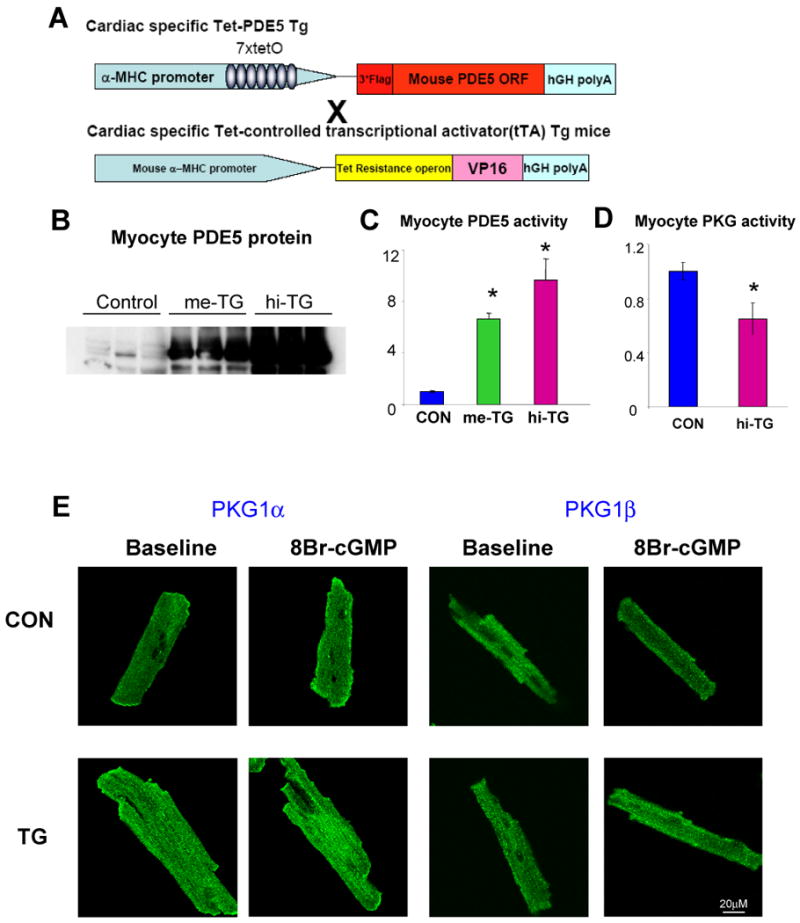
A) Schematic of cardiac specific Tet-off PDE5 Transgenic system. Two founder lines were developed. B) Western blot analysis of PDE5 expression in myocytes, C) Myocyte PDE5 activity from two founder lines (referred as me-TG and hi-TG based on expression level; *p<0.01 versus control, n=4-6 per group) D) Myocyte PKG activity of control and TG group at resting condition. *p<0.05 versus control, n=6-8 per group E) Activation of PKG1α (left) and PKG1β (right), stained green in cardiomyocyte exposed to 8Br-cGMP is reflected by rapid translocation to the plasma membrane. This occurred in control but not TG myocytes (original magnification 200×).