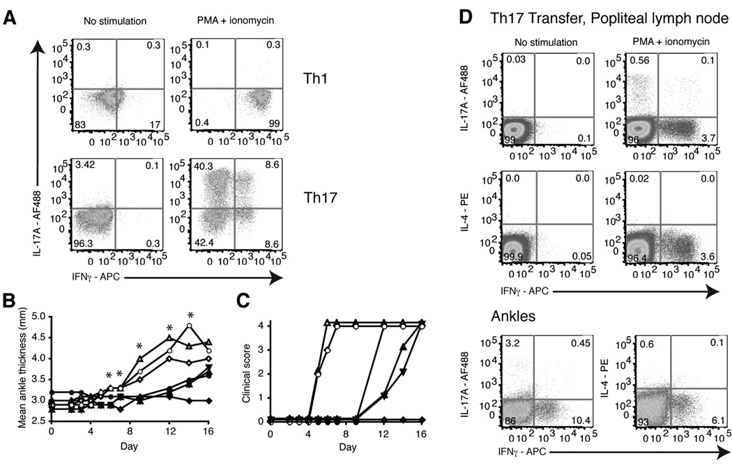
Figure 2.
Th17 and Th1 polarized KRN T cells transferred into B6.TCR.Cα−/−H-2b/g7 mice induced arthritis. Naive KRN T cells were cultured for 3 weeks under Th17 or Th1 polarizing conditions using B6.G7 splenocytes as antigen presenting cells. a, Polarized cells were stimulated for 4 h with PMA + ionomycin. T cells were gated on TCR Vβ6+ live cells and stained for intracellular IL-17A and IFNγ. b, 10×106 Th17 or Th1 polarized KRN T cells were transferred i.v. into B6.TCR.Cα−/−H-2b/g7 mice. The thickness of both rear ankles was measured as an indication of arthritis. Results are representative of at least 10 mice per group (*p < 0.05). c, Mice were examined for disease severity and given a clinical score of disease. Open symbols, Th17 cells. Filled symbols, Th1 cells. Each symbol represents one mouse. Data from three independent experiments are shown (n = 10 mice per group). Clinical score was significantly different using the Kaplan-Meir survival curves, p < 0.05 with a confidence level of 95%. d, On day 28, popliteal lymph nodes and ankles were harvested and cells stimulated for 4 h with PMA + ionomycin. T cells were gated on live Vβ6+ cells and stained for intracellular IL-17A, IFNγ and IL-4. Results are representative of 10 mice.

