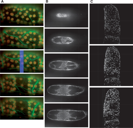
Fig. 3.
Examples of submitted movies. Each image shows a snapshot from a movie that was downloaded from PODB2. From the top to the bottom, time proceeds (A and C), or the focal plane changes from the surface to the bottom of the cell (B). These movies are available as Supplementary Movies 1, 2 and 3, respectively, at PCP online. (A) Avoidance movement of chloroplasts in a petiole cell of an Arabidopsis adult leaf expressing GFP–talin. Green and red signals show GFP-labeled actin filaments and chloroplasts, respectively. The blue rectangle in the third image represents the photo-irradiation area. (B) Serial images of microfilaments, which are visualized with GFP–ABD2, in tobacco suspension-cultured cells taken at different focal planes at metaphase during cytokinesis. (C) Stereological arrangement of GFP-labeled peroxisomes in suspension-cultured tobacco cells.

