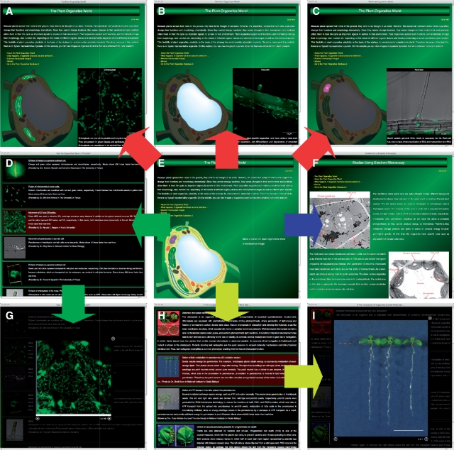
Fig. 4.
Plant Organelle World graphical user interface. On the main window (E), users can view images of organelles by moving a cursor onto each organelle in the cell, as shown by the flowchart in red. (A–C) Representative organelles, i.e. chloroplasts, a vacuole and a nucleus. From the homepage, the user can move to pages containing movies (D and G; flowchart in green), electron microscopy images (F; flowchart in blue) and images or movies showing mutant plants that have a defect in organelle function (H and I; flowchart in yellow). To view the enlarged movie or image (G and I), the user clicks on the row containing the thumbnail and description of interest on the movie (D) or mutant plant (H) pages.

