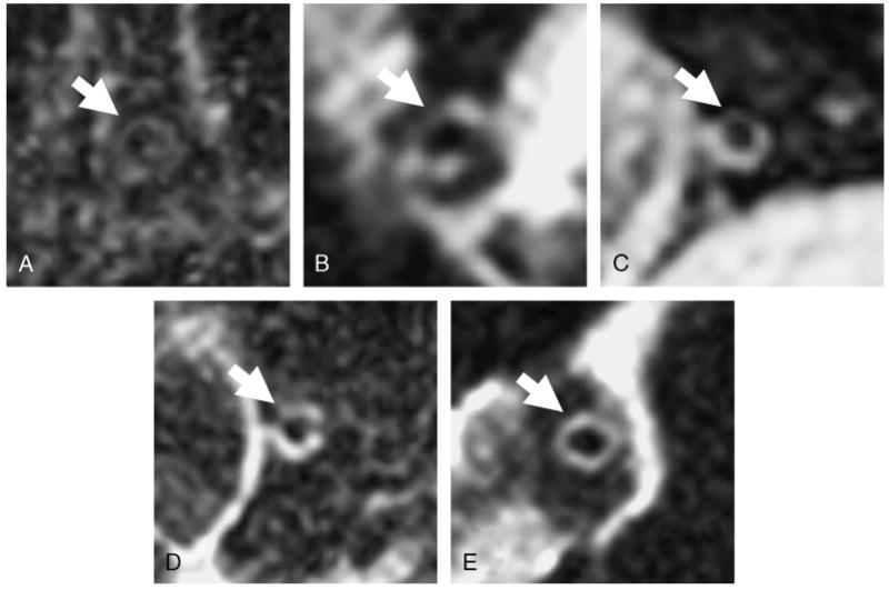
FIGURE 2.

Arteries were visually scored based on the ability of the observer to distinguish all or part of the coronary artery wall from surrounding tissue. A, A score of 1 indicated the observer was unable to distinguish the artery. B, If parts of the artery were visible (<50%) but with hazy borders, it was scored as 2. C, If at least 50% of the wall was identified and the borders were sharp, it was scored as 3. D, If only small portions of the vessel (25%) were not present with well-defined borders, it was scored as 4. E, If the entire coronary wall was visible and well defined, it was scored as 5.
