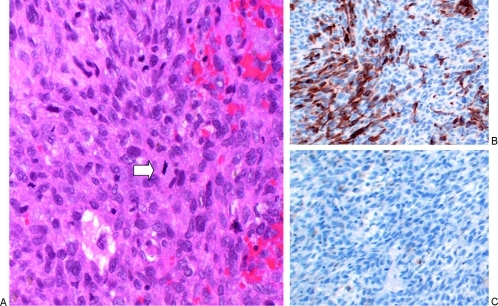
Figure 3.
Pathological findings of the postirradiation tumor. (A) A high-magnification (100×) image from a slide stained with hematoxylin and eosin shows a hypercellular neoplasm with spindle cells, pleomorphic, hyperchromatic nuclei, and mitotic figures (white arrow in A; 32 mitotic figures were found in 10 high-power fields). There is a small amount of extravasated blood. Areas of necrosis were also seen (not shown). (B) Desmin immunohistochemical stain shown at medium magnification (10×). A portion of the neoplastic cells are positive for desmin (a protein expressed in muscle). (C) S-100 immunohistochemical stain. Negative for S-100 (a protein expressed in neural crest–derived cells including Schwann cells).

