
Figure 1.
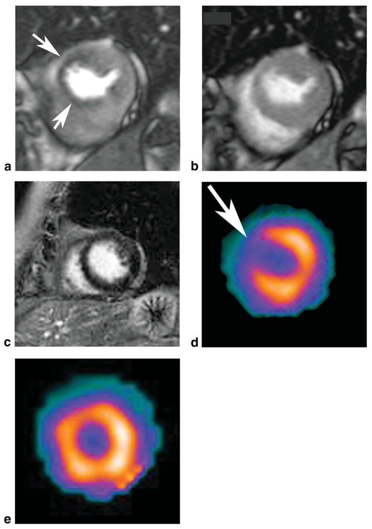
Short axis first pass adenosine stress perfusion MRI image shows a perfusion defect in the anterior-septal left ventricular wall (a, arrows), which is reversible at rest (b). Short axis delayed enhancement MR image shows no evidence of myocardial infarction (c) in this 63-year-old female patient with a high grade stenosis in the left anterior descending coronary artery on catheter directed coronary angiography. The MRI findings match the SPECT findings at stress (d) and rest (e).