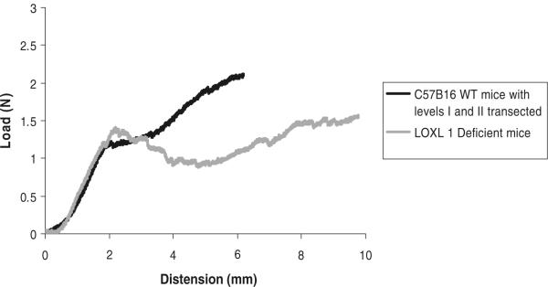
Fig. 7.
Representative load–distension curves generated following a load to failure test in WT mice after transection of level I and II vaginal supportive tissues, shown next to representative load–distension curve of LOXL1-deficient mice. Before each test, the specimens were preloaded and preconditioned

