
Fig. 2.
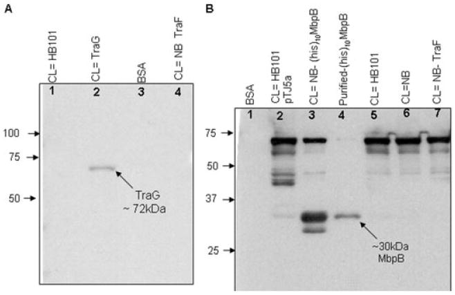
Overlay assay to determine direct TraG–MbpB interaction.
A. In vitro TraG–MbpB interaction determined by overlay with (His)10MbpB followed by detection using anti-His antibody. The blot containing cell lysate (CL) with non-His-tagged TraG (pBS140) was incubated with purified (His)10MbpB and probed with HRP-conjugate anti-His antibody. A band at the predicted size for TraG (∼72 kDa) is detected only in the lane containing overexpressed TraG (lane 2). Lane 1, CL HB101 with no plasmid; lane 2, CL with non-His-tagged TraG expressed from plasmid pBS140 (HB101); lane 3, Pure BSA; lane 4, CL with TraF expressed from plasmid pWP471 in Nova Blue (NB) cells.
B. In vitro TraG–MbpB interaction determined by overlay (reciprocal) with TraG(His)6 followed by detection using TraG antiserum. The blot containing CL with either His-tagged MbpB [pET-(His)10MbpB] or non-His-tagged MbpB (from pTJ5a) was incubated with purified TraG(His)6 and probed with TraG antiserum. Signals corresponding to ∼30–33 kDa (both types of MbpB) band were picked in three lanes containing MbpB, i.e. lane 2, CL expressing MbpB from pTJ5a; lane 3, CL expressing (His)10MbpB from pET-(His)10MbpB in NB cells; and lane 4, purified (His)10MbpB protein. No bands corresponding to this size were present in negative control lanes, i.e. lane 1, pure BSA; lanes 5 and 6, CL from HB101 and NB cells both with no plasmid; and lane 7, CL expressing TraF from pWP471, NB cells.