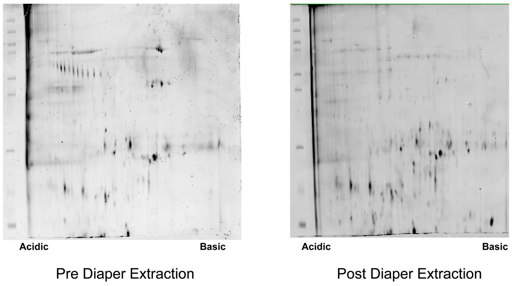
Figure 6.
Representative large format 2D gel images obtained using urine samples collected pre- and post-extraction from non-gel containing diapers. Total urine protein (150 µg) was loaded onto 18 cm IPG strips (pH 4–7 narrow range) for isoelectric focusing. Proteins were then separated in the second dimension using pre-cast 10% Duracryl-Tricine chemistry gels. Protein spots were visualized by staining the gels with SYPRO Ruby.

