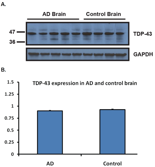
Figure 2.
TDP-43 expression in age-matched AD and control subjects. A. Five age-matched AD and four control brain samples were homogenized and run on a 10-20% Tricine Gel. The membrane was probed with the monoclonal TDP-43 antibody (EnCor Biotechnology, Gainesville, FL). One clean band migrating at the expected molecular weight was observed for all samples. The membrane was stripped and re-probed with the goat polyclonal GAPDH antibody (Santa Cruz, CA) as a loading control. B. Intensities of the signal were quantified using the NIH Image J software. There was no difference in TDP-43 expression between AD and control brain samples, after normalization to GAPDH.

