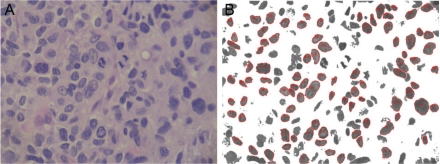
Figure 1.
Computerized nuclear morphometry. A. Osteosarcoma with large tumor nuclei before image processing. H&E, high power. B. Example of the output after image processing. The color threshold module was used to remove the background which allowed a precise selection of the nuclei. Nuclear measurements were automatically performed regarding area and roundness.

