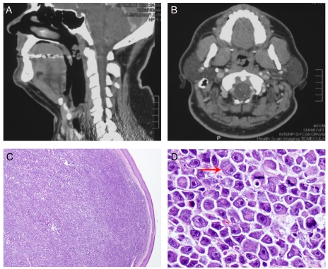
Figure 1.
Radiologic and morphologic features of the nasopharyngeal mass. A. Lateral review of the CT scan of head and neck demonstrated a posterior-lateral pedunculated nasopharyngeal mass measuring 3×2 cm; B. Anterior posterior review of the CT scan at the level of the nasopharynx showed a pedunculated mass attached to the right lateral/posterior nasopharyngeal wall; C. A diffuse dense sub-mucosal lymphoid infiltrate was seen at low magnification (H&E, original magnification 20×); D. At higher magnification, the atypical lymphoid cells showed oval to irregular nuclear contours, eccentrically located nuclei, prominent nucleoli, and abundant cytoplasm with frequent peri-nuclear hofs. Brisk of mitotic rate was easily appreciated (H&E, original magnification 400×).

