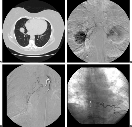
Figure 2.
(A) Computed tomography scan showing lung metastasis in a patient with known renal cell carcinoma and massive hemoptysis. Bronchoscopy localized bleeding to the right side. (B) Bronchial angiogram was performed with selective catheterization of an intercostal bronchial trunk, which gives rise to right and left bronchial arteries as well as right intercostals. Demonstrated is enhancement of a large right lung lesion and partial supply to a left lung lesion. (C) Postembolization angiogram was performed demonstrating stasis of antegrade flow to the right bronchial artery. Embolization was performed via a coaxial system (Renegade Hi-Flo microcatheter [Boston Scientific, Natick, MA] through a Mikaelsson catheter [Cook Medical, Bloomington, IN]) utilizing polyvinyl alcohol particles of 355 to 500 microns. (D) During bronchial angiography, recognition of spinal cord supply via the anterior spinal artery, or artery of Adamkiewicz (arrowheads), is critical to avoid nontarget embolization leading to cord ischemia.

