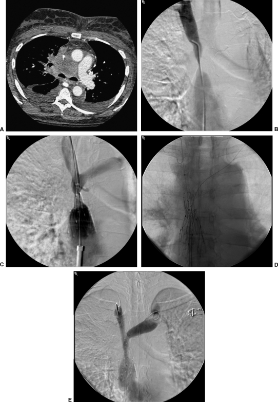
Figure 3.
(A) Computed tomography scan of a patient with metastatic bronchogenic carcinoma. The superior vena cava (SVC) is encased in this patient with dyspnea and facial swelling. (B) SVC gram demonstrates severe stenosis of the SVC from external compression. (C) SVC gram during deployment of a 25-mm diameter Gianturco Z stent (off-label use; Cook Medical, Bloomington, IN). (D) Self-expanding kissing brachiocephalic vein stents were utilized to preserve the venous confluence. (E) Completion venogram demonstrating technical improvement in SVC patency. This patient's symptoms were significantly improved within 48 hours.

