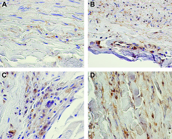
Figure 4.
Immunohistochemical staining of PEPD demonstrates expression in both aneurysmal and non-aneurysmal aortic tissue. Immunohistochemical staining using a commercially available specific antibody against PEPD was performed on formalin-fixed paraffin embedded tissue sections of non-aneurysmal abdominal aorta (A, B) and AAA (C, D) with staining observed in medial (A) and adventitial layers of controls and throughout the aneurysmal wall. Negative control staining with non-immune serum showed no staining (data not shown). Regions of positive staining appear reddish-brown with hematoxylin counterstaining in blue.

