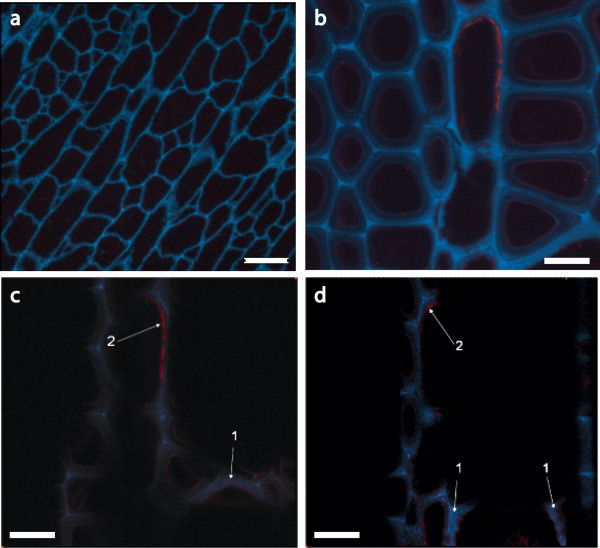
Figure 9.
Localization of E1 in transgenic tobacco shown in confocal microscopic images of sectioned WT and E1-containing tobacco. All images were obtained by using immunofluorescence confocal laser microscopy. An E1cd primary antibody and an Alexa Fluor 488 antimouse secondary antibody were used and spectrally deconvoluted to show antibody in red and nonspecific autofluoresence in blue. (a) Negative control WT tobacco confocal image stack at ×600 magnification. Note the absence of E1 antibody staining in this image (red). (b) E1cd-transformed tobacco image stack at ×1,200 magnification. (c and d) Individual sections of the confocal image stack presented in (b) illustrate that E1cd antibody stained E1cd-transformed tobacco. Arrows indicate (1) deconvoluted E1 antibody signal present within a tobacco cell wall section (red) and (2) E1 antibody staining present on the inside of a cell wall (red). Blue coloring denotes the residual plant autofluorescence signal. Scale bar in (a), 25 μm; original magnification ×600. Scale bars in (b-d), 50 μm; original magnification, ×1,200.

