Figure 4.


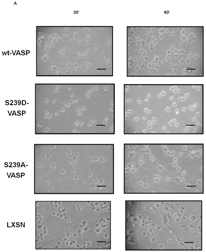
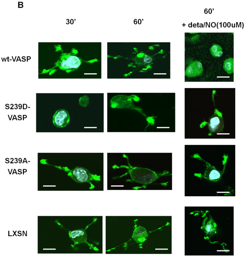
NO and expression of S239D-VASP inhibit membrane protrusion formation and SMC spreading within collagen. (A) Cell morphology was analyzed by phase contrast microscopy 30 and 60 minutes after the cells were seeded within collagen. Scale bar represents 10μm. (B) wt-VASP, LXSN, S239A-VASP or S239D-VASP SMCs were pretreated with or without 100 μM deta NONOate for 30 minutes at room temperature and then seeded within collagen gel in presence or absence of deta NONOate. After 30 and 60 minutes, the cells were analyzed by fluorescence microscopy after staining with TRITC-conjugated phalloidin. Scale bar represents 5μm. The photomicrographs are representative of three independent experiments.
