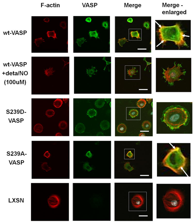
Figure 5.

Colocalization of VASP and F-actin is inhibited by phosphorylation of VASP serine239. wt-VASP, LXSN, S239A-VASP or S239D-VASP SMCs were plated on polymeric collagen-coated glass slides, and wt-VASP SMCs were pretreated with 100 μM deta NONOate for 30 minutes at room temperature and then plated on polymeric collagen-coated glass slides in the presence of 100 μM deta NONOate. 3 hours after plating localization of VASP and F-actin was analyzed by staining with rhodamine-phalloidin (red) and VASP (green). Boxed regions are enlarged in insets. Arrows indicated where VASP and F-actin are colocalized (yellow). Scale bar represents 5μm.
