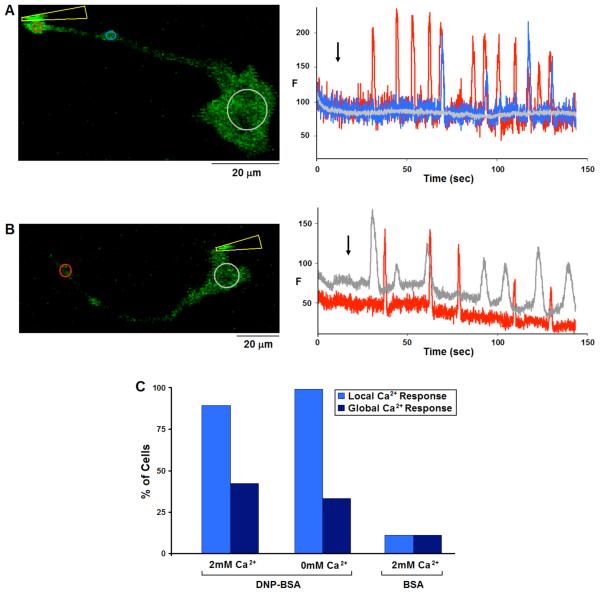
Figure 4. Stimulation of RBL cells by contact with Ag-coated micropipette.
A and B) Representative cells expressing GCaMP2, sensitized with IgE and stimulated with DNP-BSA conjugated micropipettes. A) Micropipette (indicated by yellow arrowhead) contacting the cell at the tip of a protrusion elicits a train of spatially restricted Ca2+ puffs, each traveling no more than 30μm along the protrusion. B) Contact stimulation at the cell body results repetitive Ca2+ puffs in the cell body that sometimes propagate as a wave to the protrusion. Left panels show images with ROIs defined; Right panels show Ca2+ concentration changes in ROIs of corresponding color. Black arrow indicates initiation of contact between micropipette and cell. C) Histogram showing percentage of cells responding with local Ca2+ puffs only (light blue) or more global Ca2+ elevation (dark blue) due to contact with DNP-BSA-conjugated micropipettes in the presence (n=28) or absence (n=18) of extracellular Ca2+, or with unmodified BSA-conjugated micropipettes (n=18).

