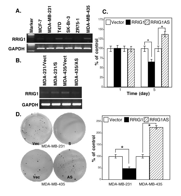
Figure 2.
RRIG1 expression and modulation of breast cancer cell growth and colony formation. All experiments were repeated once with similar results. A, Semiquantitative RT-PCR. Breast cancer cell lines were grown in monolayer, and RNA was then isolated from the cells and subjected to semiquantitative RT-PCR analysis of RRIG1 expression. B, Semiquantitative RT-PCR.pCDNA3.1 carrying RRIG1 sense and antisense cDNA was transiently transfected into MDA-MB-231 and MDA-MD-435 cells, respectively. The vector-only plasmid was used as a control. The cells were grown in G418-containing medium, and RNA from the cells was isolated and subjected to semiquantitative RT-PCR analysis. C, Cell viability assay. The gene-transfected cells were grown in G418-containing medium for 1 or 5 days, and cell viability was measured using the MTT assay. D, Colony formation assay. The gene-transfected cells were grown in soft agar with G418-containing medium for 21 days, and cell colonies were then visualized by incubation with MTT, counted, and summarized. *p < 0.05 vs. the control.

