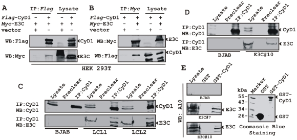
Figure 3. EBNA3C forms a complex with Cyclin D1 in human cells.
A-B) 15 million HEK 293T cells were co-transfected with myc-tagged EBNA3C and flag-tagged Cyclin D1 vectors. In each case control samples were balanced with empty vector. Cells were harvested at 36 h post-transfection and approximately 5% of the lysed cells were saved as input and the residual lysate was immunoprecipitated (IP) with 1 µg of indicated antibody. Lysates and IP complexes were resolved by 10% SDS-PAGE and western blotted (WB) with the indicated antibodies. C) 50 million BJAB cells and two different clones of EBV transformed lymphoblastoid cell lines - LCL1 and LCL2, and in D) BJAB cells stably expressing EBNA3C (BJAB_E3C#10) along with BJAB control cells were collected and lysed in RIPA buffer. Protein complexes were immunoprecipitated with Cyclin D1 specific antibody and samples were resolved by a 10% SDS-PAGE followed by western blot with antibodies as indicated. E) Either GST control or GST-cyclin D1 beads were incubated with lysates prepared from 50 million BJAB cells and two different clones of BJAB cells stably expressing EBNA3C (BJAB_E3C#7 and #10). EBNA3C was detected by western blot with the specific monoclonal antibody (A10). Coomassie staining of a 12% SDS-PAGE resolved purified GST and GST-Cyclin D1 proteins used in this study is shown in the right panel.

