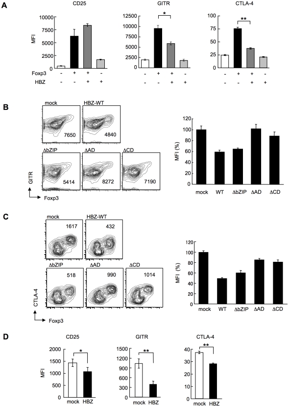
Figure 6. HBZ inhibites Foxp3-mediated CTLA-4 and GITR expression in vitro.
(A) Mouse CD4+CD25− T cells co-transduced with the retroviral vectors were stained with the indicated antibodies. Mean fluorescence intensity (MFI) of CD25, GITR, and CTLA-4 in GFP/NGFR double-positive cells are shown as mean ± SD. for triplicate culture. *, P<0.01; **, P<0.001 by two-tailed Student t-test. (B and C) CD4+CD25− T cells transduced with the pMXs-Ig vector encoding wild-type or mutant HBZ, and pGCSamIN-Foxp3 vector were stained with anti-GITR (B) or anti-CTLA-4 (C) antibody in addition to anti-NGFR antibody, and then analyzed by flow cytometry. Left, numbers in density plots indicate MFI of GITR (B) or CTLA-4 (C) in GFP/NGFR double-positive cells. Representative data from three independent experiments are shown. Right, relative MFI of wild type or mutated HBZ compared to mock transduced cells was shown as mean ± SD (n = 3). (D) HBZ transduction in Foxp3+ Treg cells inhibited the endogenous expression of Treg associated molecules. Mean fluorescence intensity (MFI) of CD25, GITR, and CTLA-4 in CD4+Foxp3+NGFR+ cells are shown as mean ± SD. for triplicate culture. *, P<0.05; **, P<0.01 by two-tailed Student t-test.

