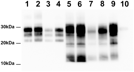
Figure 1. PrPSc Western Blot detection in sheep and tg338 mice inoculated with Atypical/Nor98 scrapie and classical scrapie tissues (10% tissue homogenate).
Lane 1: posterior brainstem from a Langlade classical scrapie affected sheep (case 10). Lane 2: brain from a tg338 mouse inoculated with striated muscle (semi-membranous) from the same sheep (case 10). Lane 3: posterior brainstem from a PG127 classical scrapie orally inoculated sheep (case 12). Lane 4: brain from a tg338 mouse inoculated with striated muscle (extra-ocular motor muscle) from the same PG127 classical scrapie affected sheep (case 12). Lane 5: cerebral cortex from an AFRQ/AFRQ Atypical/Nor98 scrapie natural case (case 1). Lane 6: AHQ/AHQ sheep (cerebellum-case 9) intra-cerebrally challenged with natural atypical scrapie (case 1). Lane 7 to 9: brain homogenates from tg338 mice inoculated with peripheral tissues from atypical scrapie cases. Lane 7: retropharyngeal lymph node (case 2). Lane 8: sciatic nerve (case 9). Lane 9: striated muscle (case 8). No PrPSc was observed in control tg338 mice inoculated with retropharyngeal LN from a negative control sheep (Lane 10).

