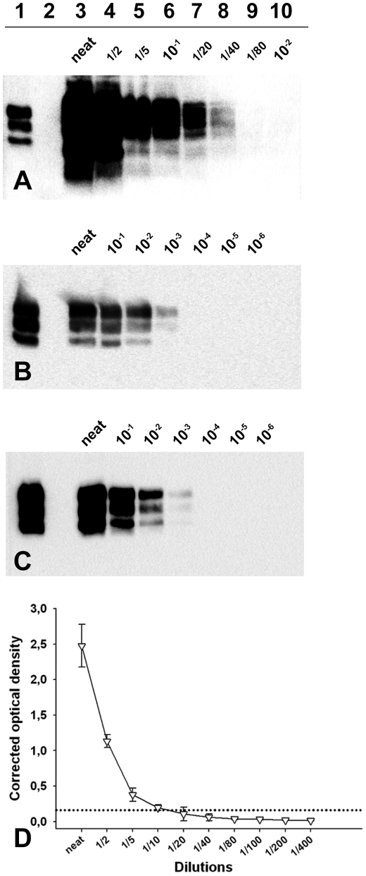
Figure 3. PrPSc detection limit of a Langlade classical scrapie isolate, a PG127 classical scrapie isolate and an Atypical/Nor98 scrapie isolate.
Dilution series from (A, D) Atypical/Nor98 scrapie isolate (case 9- cerebral cortex), (B) Langlade–case 10- posterior brainstem) and (C) PG127 (case 12- posterior brainstem) classical scrapie isolate, that were prepared in negative sheep brain homogenate. The tissue homogenates (see methods) are the same than the one used for endpoint titration in tg338 mice (Table 3). The samples were processed for PrPSc Western-blotting (A–C, TeSeE WB kit – BioRad, anti-PrP SHa31 antibody). After extraction (25 mg of brain equivalent material), the re-suspended pellet was either entirely loaded on lanes (A: all lanes – B and C: Lanes 4–6) or diluted in Laemmli's buffer before loading (B: lane 3, 1/50 – Lane 4, 1/10 – C: lane 3, 1/20). These dilutions were necessary to avoid a saturation of the signal. A classical scrapie control was included on the three gels to calibrate the signal (Lane 1). (D) The atypical/Nor98 scrapie isolate dilution serie (case 9: cerebral cortex) was tested using a commercially available rapid TSE test (TeSeE Sheep and Goat - BioRad) used for field TSE screening in small ruminants. Three different aliquots of each dilution were independently tested. The extractions were carried out. Results are presented as the mean +/− SD corrected optical density values. The cut off value (0.162 OD– dotted lines) was established as the mean of four negative control optical densities + 0.150 OD.

