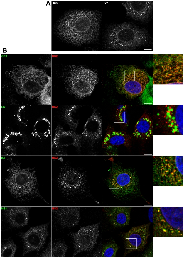
Figure 3. Subcellular localization of NS2.
(A) NS2 accumulates in dotted structures. Huh-7 cells electroporated with JFH-HA RNA were grown on coverslips and fixed at 48h and 72h post-electroporation. The subcellular localization of HA-NS2 was analyzed by immunofluorescence using an anti-HA antibody. (B) Colocalization of NS2 dotted structures with cellular and viral markers. JFH-HA electroporated cells grown on coverslips were fixed at 72h post-electroporation and processed for double-label immunofluorescence for HA-NS2 (red) and the ER marker calreticulin (CRT in green) or the LDs stained with BODIPY 493/503 (green). JFH-HA electroporated cells were further stained for HA-NS2 (red) and HCV proteins NS3 or E2 (green). The nuclei were stained with DAPI. Representative confocal images of individual cells are shown in grey and the merge images in color. Zoomed views of the indicated areas are shown in the right column. Bar, 10 µm.

