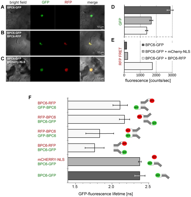
Figure 6. AtBPC6 forms parallel dimers in planta.
(A–C) Confocal laser scanning microscopy analysis of GFP-/RFP-fusion proteins and mCherry-NLS in transiently co-transformed Nicotiana benthamiana epidermis cells. All proteins localize to the nucleus. GFP fluorescence intensities (D) and RFP-FRET fluorescence intensities (E) under GFP-excitation light. (F) In vivo GFP fluorescence life time measurements of all four possible GFP/RFP protein combinations fused to either the N- or the C-terminus of AtBPC6. BPC6-GFP and BPC6-GFP/mCherry-NLS combinations serve as controls. Pictographs (right hand side) display the only possible zipper orientations that are in accordance with the GFP fluorescence life time measurements.

