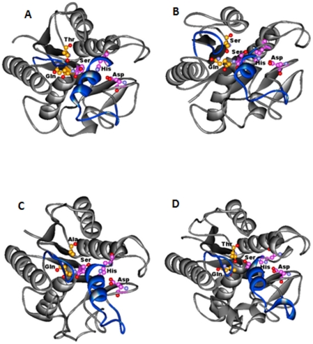
Figure 3. The top view of the serine hydrolases indicating a closed conformation of the lid insertion on the active site.
(A) Rv1430 PE-PPE domain. (B) Rv1800 PE-PPE domain. (C) Rv1184c PE-PPE domain. (D) PDB_ID: 3AJA. The protein is represented in grey, the lid insertion in blue, the side chains of the amino acids in the catalytic triad and oxyanion hole are indicated in ball and stick.

