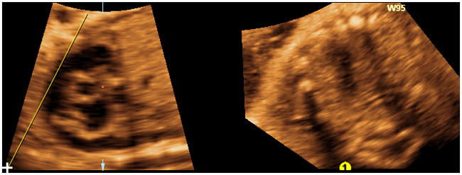
Figure 6. “ Swing technique” (placement of “swing” line).

At the top of the longitudinal view of the ductal arch image, the “swing” line is begun approximately above the center of the right ventricle and is fixed (but not locked) on this end only. The line is drawn from the top to the lower left hand corner of the image, making sure it is lateral to the ductal arch. The line should remain unlocked with the cursor (not the “lollipop”) visualized at its inferior end.
