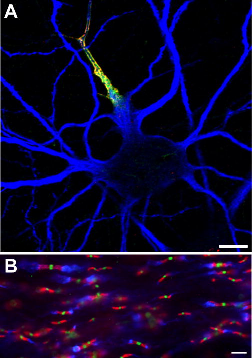
Figure 1.
Ion channels are clustered in high density at multiple sites along axons. A, Axon initial segment (AIS) of a cultured hippocampal neuron immunolabeled using antibodies against Nav channels (red), ankG (green), and the microtubule associated protein 2 (MAP2) to define the somatodendritic domain (blue). B, Central nervous system nodes of Ranvier immunolabled using antibodies against Nav channels (green), Caspr (red), and Kv1.2 K+ channels (blue). Scale bars, A = 10 μm; B = 5 μm.

