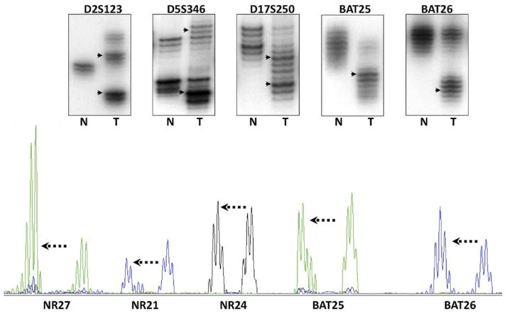
Figure 3.
MSI was initially discovered by autoradiography analyses of the PCR products after separation by gel electrophoresis (upper panel). N refers to DNA from the normal colon, and T from the tumor. The DNA polymerase used in PCR also has difficulty with the accurate amplification of templates, which is thought to be the explanation for the “ladder” of DNA bands that can be seen in the lanes for normal and tumor DNA. The upper panel illustrates the use of the 5 markers recommended by the National Cancer Institute consensus group; these consist of 3 dinucleotide repeats and 2 mononucleotide repeats (BAT25 and BAT26). In each instance, the DNA in the tumor has undergone somatic mutations (frequently, but not always, deletions), and the PCR product migrates to a different position on the gel, as indicated by the arrowheads. The lower panel shows the PCR products as they are analyzed by most laboratories using automated DNA sequencing with fluorescent primers. In this instance, 5 mononucleotide repeats have been analyzed, and in each instance, the mutations consist of deletions with different electrophoretic mobility (mutant alleles indicated by the arrows).

