
Figure 4.
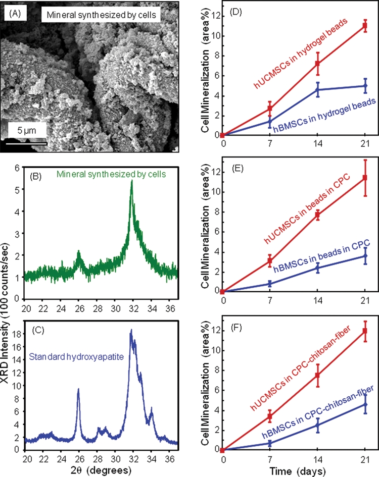
hUCMSC and hBMSC mineralization. (A) SEM of minerals synthesized by the encapsulated hBMSCs at 21 days. (B) XRD of minerals by hBMSC at 21 days. (C) XRD of a known hydroxyapatite formed at 37°C, provided by Dr. Shozo Takagi at NIST. The XRD patterns confirm that the substance made by the cells was apatitic. (D-F) Comparison of mineralization by hUCMSCs and hBMSCs (mean ± SD; n = 5). hUCMSCs synthesized more bone minerals than hBMSCs while encapsulated in all 3 constructs (p < 0.05). Mineralization increased rapidly from 7 to 21 days (p < 0.05).