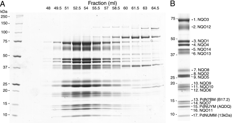
FIGURE 2.
Subunit composition of complex I from P. denitrificans. A, SDS-PAGE analysis of Superdex 200 fractions. Protein was stained with Coomassie Blue. Molecular mass markers are in the 1st lane and are indicated at the left. B, purified enzyme, as used for MS analyses. Identified subunits are indicated on the right. Hydrophobic subunit Nqo11 is weakly stained by Coomassie Blue but is more clearly visible in silver-stained gels (not shown). Positions of Mr markers are shown on the left.

