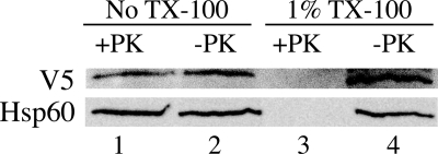
FIGURE 6.
Proteinase K digestion of CHO cell mitochondria expressing epitope-tagged rat MTHFD2L. Mitoplasts were prepared by the digitonin method and treated with 100 μg/ml proteinase K (PK) as described under “Experimental Procedures.” Protease treatment was stopped by the addition of PMSF to 1 mm, and the mitoplast suspensions were centrifuged through sucrose cushions. The resulting pellets were resuspended in equal volumes of HMS without BSA, fractionated on a 10% SDS-polyacrylamide gel, and subjected to immunoblotting with anti-V5 (1:1500 dilution) or anti-Hsp60 (inner membrane/matrix marker; 1:1200 dilution) antibodies. Mitoplasts incubated with 1% Triton X-100 (TX-100) in the presence or absence of proteinase K were used as controls. These Triton X-100-treated controls were analyzed directly without centrifugation through sucrose cushions. MTHFD2L migrated at an apparent molecular mass of 37 kDa, and Hsp60 migrated at an apparent molecular mass of 60 kDa.

