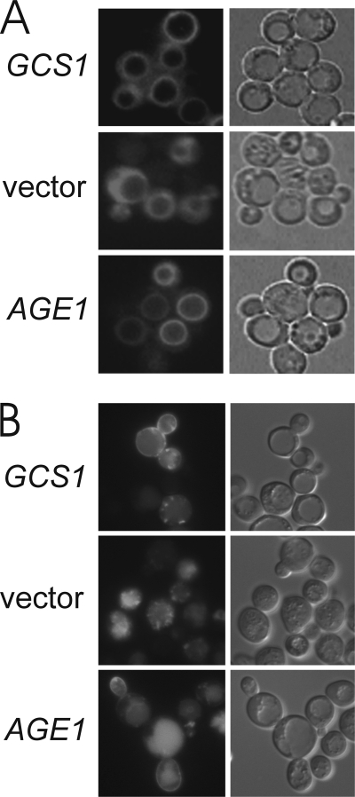
FIGURE 2.
Increased AGE1 dosage restores post-Golgi function in age2Δ gcs1 cells. Cells were visualized by fluorescence (left panels) and differential interference contrast microscopy (right panels). A, mutant age2Δ gcs1-3 cells growing at 30 °C and carrying plasmid-borne GCS1 or AGE1 genes were stained with FM 4-64 as described (14) and then incubated in fresh medium at 37 °C for 45 min before visualization. B, mutant age2Δ gcs1-4 cells carrying p416-GFP-Snc1 and plasmid-borne GCS1 or AGE1 genes were grown at 23 °C and then incubated at 37 °C for 3 h before visualization.

