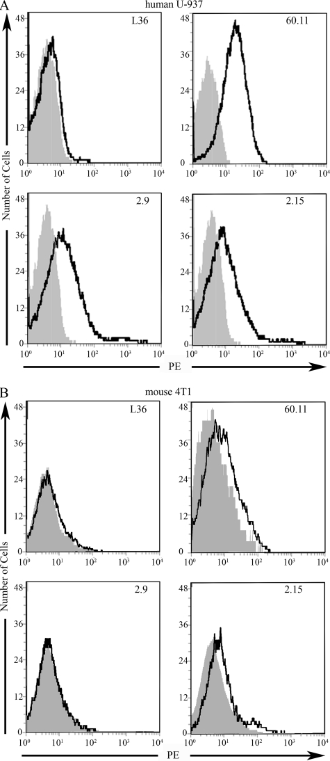
FIGURE 2.
Reactivity of scFv fragments to cell surface expressed p32. A, binding of anti-p32 mAb and purified scFv fragments to phorbol myristic acid-stimulated human U-937 cells by flow cytometry. B, binding of anti-p32 mAb and purified scFv fragments to mouse 4T1 cells by flow cytometry. For purified scFv fragments, the bound scFv was detected with sequential incubations with 9E10 anti-Myc mAb and phycoerythrin (PE)-labeled goat anti-mouse IgG. FACScan histograms show the binding of each scFv clone (bold line) and the backgrounds of phycoerythrin-conjugated secondary antibodies (gray).

