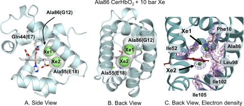
FIGURE 1.
Effects of 10 atm of xenon on the structure of L86A CerHbO2 (PDB code 2xkh). A, side view looking through the heme propionates into the protein interior. B, back view looking between the E and H helices through the apolar tunnel. The empty space in the tunnel is highlighted in gray. The heme group, proximal His, and Gln-44(E7) are shown as sticks with O atoms in red, N atoms in blue, and C atoms in white. The xenon atoms are shown as green spheres at half their van der Waals radii to indicate partial occupancy and designated as Xe1 and Xe2. The Ala-86(G12) and Ala-55(E18) methyl side chains are shown as green sticks. C, electron density map of the amino acids and xenon atoms lining the tunnel using the same orientation as in panel B are shown. Amino acid side chains are shown as ball and stick models, and the xenon atoms are drawn as green spheres. The electron densities (2Fo − Fc map contoured at 1σ) are shown in magenta for the side chains and in blue for the xenon atoms.

