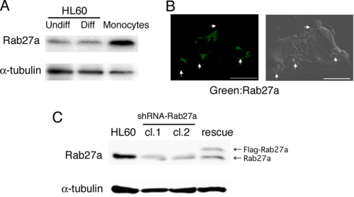
FIGURE 1.
Rab27a is expressed in human phagocytic cells. A, Rab27a expression in HL-60 cells, macrophage-like differentiated (Diff) HL-60 cells treated with vitamin D3 and 12-O-tetradecanoylphorbol-13-acetate for 3 days, and peripheral blood-derived monocytes were analyzed by immunoblotting with antibodies against human Rab27a and α-tubulin as a loading control. B, a phagocytosis assay was performed using macrophage-like differentiated HL-60 cells and C3bi-zymosan for 30 min as described under “Experimental Procedures.” Distribution of Rab27a was analyzed by staining with anti-Rab27a antibodies (left) and a differential interference contrast image (right). Scale bar = 5 μm. Arrowheads show C3bi-zymosan particles. C, decreased expression of Rab27a by Rab27a shRNA transfer into HL-60 cells (clones 1 (cl.1) and 2 (cl.2)) and its recovery by FLAG-rescue-Rab27a into Rab27a knockdown cells by immunoblotting with antibodies against human Rab27a and α-tubulin as a loading control.

