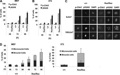
FIGURE 5.
TopBP1 ablation activates DNA damage signaling. Cre-retrovirus infected MEF (A) and 3T3 cells (B) were immunostained with anti-p-Chk2 (Thr68) or anti-γH2AX (Ser139) antibody at the indicated times after viral infection (supplemental Fig. S4). Cells found to be positive for p-Chk2, and γH2AX signaling was counted and graphed. Three independent experiments were performed with 300 cells counted at least for each experiment. C, the indicated 3T3 cells were treated with UV (100 J/m2) 96 h after viral infection and then immunostained 3 h after the irradiation. D, at the indicated times (MEF) or 96 h (3T3) after viral infection, the nuclear morphologies were visualized by DAPI staining. The percentages of cells containing abnormal nuclear morphology (binuclear and micronuclear cells) are indicated. Three independent experiments using at least 100 cells for each experiment were averaged.

