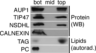
FIGURE 4.
AUP1 localizes to the LD fraction. A431 cells were labeled with radioactive oleic acid and fractionated in a sucrose gradient. Isolated LDs (top), middle (mid) and bottom (bot) fraction, were analyzed by Western blotting (WB). AUP1 localizes, similar to the LD marker proteins NSDHL and TIP47, to the LD and the bottom fraction. In contrast, the ER membrane protein calnexin was not detected in the LD fraction. The purity of the LD preparation was validated by the presence of radioactive triacylglycerol (TAG) and the absence of radioactive phosphatidylcholine (PC).

