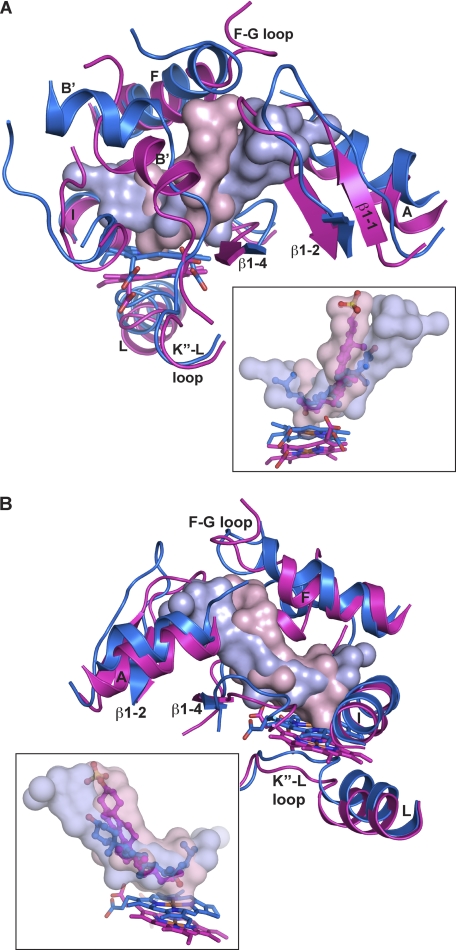
FIGURE 4.
Superimposed views of the active sites in CYP11A1 and CYP46A1 illustrating positioning of the secondary structural elements (A and B) and sterol substrates (insets). Secondary structural elements and the heme group are colored in marine in CYP11A1 and magenta in CYP46A1. The solvent-occupied volume of the active site is shown either as a solid or semitransparent surface in light blue in CYP11A1 and light pink in CYP46A1. 22HC is in blue, and cholesterol 3-sulfate is in magenta. Coloring of atoms is the same as in Fig. 1.

