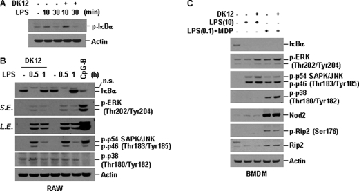
FIGURE 4.
DFK1012 does not block phosphorylation and degradation of IκBα and activation of MAP kinase by LPS or MDP stimulation. A and B, RAW264.7 cells were pretreated with DFK1012 (DK12) for 1 h and then stimulated with LPS (10 ng/ml) for the indicated periods in the presence or absence of DFK1012. Cells were treated with CpG-B (1 μm) for 30 min as a positive control. Total cellular extracts were subjected to Western blot analysis for phosphorylated IκBα (p-IκBα), IκBα, phosphorylated ERK (p-ERK), phosphorylated p54 and p46 SAPK/JNK (p-SAPK/JNK), phosphorylated p38 (p-p38), and actin. n.s., nonspecific; L.E., long exposure; S.E., short exposure. C, BMDM were pretreated with or without DFK1012 for 1 h and then stimulated with LPS (10 ng/ml) for 30 min (lanes 1–3). Cells were pretreated with LPS (0.1 ng/ml) for 5 h and then stimulated with MDP (100 μg/ml) for 30 min in the presence or absence of DFK1012 (lanes 4 and 5) Total cellular extracts were subjected to Western blot analysis for IκBα, p-ERK, p-SAPK/JNK, p-p38, Nod2, phosphorylated Rip2 (p-Rip2), total Rip2, and actin. Data are representative of three independent experiments with similar results.

