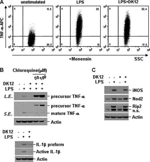
FIGURE 7.
DFK1012 triggers post-translational degradation of TNF-α through the lysosomal pathway. A, RAW264.7 cells were treated with LPS (10 ng/ml) in the presence or absence of DFK1012 (DK12) for 8 h in the presence of monensin (2 μm). The expression of intracellular TNF-α was analyzed by flow cytometry. APC, allophycocyanin. B, RAW cells were stimulated with LPS in the presence or absence of DFK1012 or chloroquine (50 and 100 μm) for 12 h. The expression of intracellular TNF-α or IL-1β was analyzed by Western blot analysis. n.s., nonspecific; L.E., long exposure; S.E., short exposure. C, RAW264.7 cells were stimulated with LPS (10 ng/ml) in the presence or absence of DFK1012 for 18 h. Total cellular extracts were subjected to Western blot analysis for iNOS, Nod2, Rip2, and actin. Data are representative of three independent experiments with similar results.

