FIGURE 5.
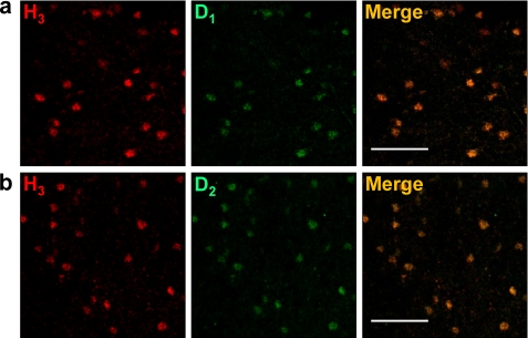
Co-localization between H3R and D1R or D2R in striatal MSNs. Confocal microscope representative images of coronal sections from striatal slices are shown. Slices were labeled with anti-H3R antibody (red). Labeling (green) using an anti-D1R antibody (a) or an anti-D2R antibody (b) is also shown. In a and b, colocalization is shown in yellow. Scale bars, 60 μm.

