FIGURE 7.
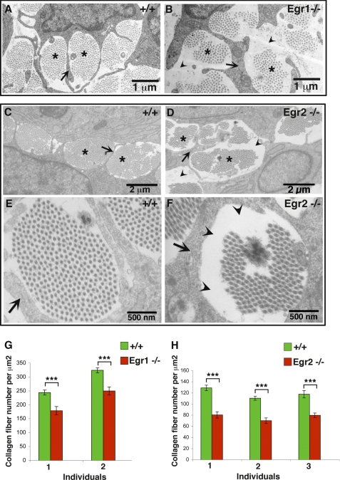
Collagen fibril organization in Egr1- and Egr2-deficient tendons. Transmission electronic microscopy images of wild type (A, C, and E), Egr1−/− (B), or Egr2−/− (D and F) hind limb tendons at E18.5. Collagen fibrils are organized in bundles (asterisks in A–D) surrounded by cytoplasmic processes of tenocytes (arrows in A–F). E and F, show high magnification of a fibril bundle in control (E) and Egr2-defective tendon (F). Collagen fibrils are less abundant in mutants (B, D, and F) compared with control tendons (A, C, and E), leaving some unoccupied spaces (arrowheads in B, D, and F). G and H, the number of collagen fibrils per surface unit was analyzed in each sample from different mouse groups. The number of collagen fibril per surface unit diminished in the absence of Egr1 and Egr2 activity compared with control tendons. Histograms are expressed as means and S.E. Asterisks in the histograms indicate p values ***, p <0.001.

