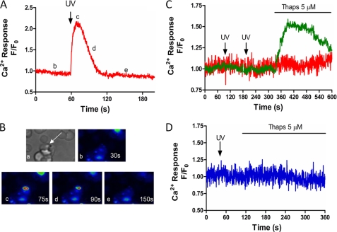
FIGURE 1.
Flash photolysis of caged-IP3 induces calcium release in P. falciparum-infected RBC. A and B, P. falciparum-infected RBCs were loaded in HBSS with Fluo4-AM (5 μm) and caged-IP3 (2 μm) for 45 min, then allowed to adhere to poly-l-lysine-coated coverslips. Changes in intracellular Ca2+ were monitored at 2 Hz using a spinning disc confocal microscope coupled to a CCD camera. Flash photolysis of caged-IP3 was achieved with a nitrogen-charged UV laser. A, representative trace of UV-induced Ca2+ increase in intact P. falciparum (UV flash indicated by arrow at 60 s). B, confocal images of the cell in Panel A to show: (a) transmitted light image depicting P. falciparum within RBC (arrow); (b–e) changes in Ca2+ are shown in pseudocolor (blue lowest and red highest [Ca2+]) at (b) baseline (t = 30 s) (c) peak Ca2+ transient (t = 75 s), (d) half-peak height (t = 90 s), and (e) return to baseline (t = 150 s). Data are representative of 81 cells from 15 experiments. C, representative traces of infected (green) and uninfected (red) RBC loaded with Fluo4-AM in the absence of caged-IP3 (UV flashes at 75 and 180 s). D, representative trace of uninfected RBC in the presence of caged-IP3 (UV flash at 40 s). Thapsigargin (5 μm, Thaps) was added as indicated.

