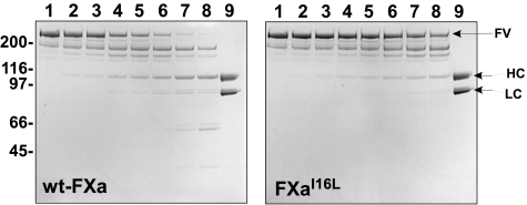
Figure 2.
Activation of FV by membrane-bound FXa. Plasma-derived FV (400nM) was incubated with increasing concentrations of wt-FXa or FXaI16L in the presence of PCPS (50μM) for 10 minutes at 25°C. Samples (4 μg/lane) were then resolved on sodium dodecyl sulfate polyacrylamide gel electrophoresis and visualized by staining with Coomassie Blue R-250. Lane 1, platelet-derived (pd)-FV, no FXa; lanes 2-8, FXa (wt or variant) at 0.5, 1.0, 5.0, 10, 20, 50, and 100 nM; lane 9, recombinant FVa. The apparent molecular weights of the standards are indicated on the left. HC, heavy chain; LC, light chain. The data are representative of 2 similar experiments.

