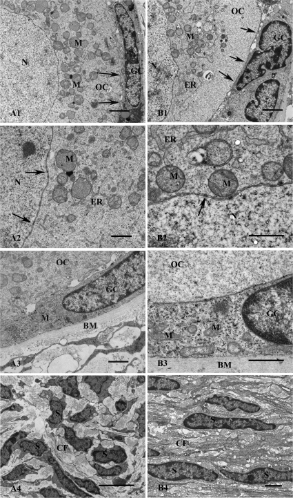
Figure 6.
Ultrastructure of follicles within human ovarian tissue after culture for 24 h in non-vitrified (A) and vitrified (B) groups. The oocyte nuclei (N) surrounded by nuclear membrane in a primordial follicle is shown (A1). Cytoplasm contains well-defined M. The intact contact between GC and OC is seen (arrows). The N and cytoplasmic organelles, M and ER are well defined. The nuclear pores (arrows) are clearly seen (A2). The contact between GC, OC and BM is shown (A3). GC contains hetero- and euchromatin in their nuclear. Well-defined M scattered in the cytoplasm are seen (A3). Image of stromal tissue composing bundles of CF and S (A4). Scale bars; A1 = 2 µm, A2–A3 = 1 µm and A4 = 5 µm. Overview of a primary follicle with OC and GC showing well-defined M and ER and intact contact between GC and the OC (arrows) (B1). Detail of the border between N and cytoplasm in OC showing well-defined nuclear membrane (arrows), ER and M containing cristae (B2). Granulosa cell in contact with OC and BM is shown (B3). Numerous M are scattered in the cytoplasm of the GC (B3). A sample of stroma containing numerous CF and flattened S is shown (B4). Scale bars; B1 = 2 µm, B2–B3 = 1 µm and B4 = 2 µm.

