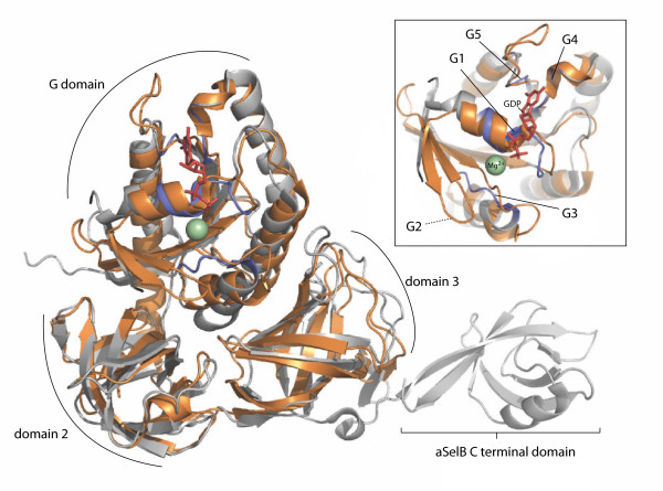
Figure 3.
Structural alignment of aSelBL and aSelB. aSelBL ("EF-Pyl") from Methanosarcina mazei (PDB ID 2ELF) is shown in bronze, and aSelB from Methanococcus maripaludis in complex with GDP (PDB 1WB1) is shown in silver. Blue colouring indicates the location of the nucleotide binding motifs in aSelB. GDP and the magnesium ion bound to aSelB are shown as red sticks and a green sphere respectively. The inset box shows a more detailed view of the G domain, with the G1 and G3-5 motifs from aSelB labelled. As G2 is disordered in aSelB, the equivalent region in aSelBL is indicated.

