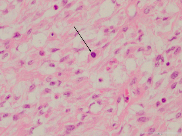
Figure 6.

Phyllodes tissue H&E staining (×40 magnification). High-power view shows the cellularity of the stroma, with presence of mitosis (arrow) and moderate cellular pleomorphism. The presence of increased mitotic activity (five per 10 high-powered fields (hpfs) in this patient), as well as the moderate cellular pleomorphism, indicates a diagnosis of "borderline" phyllodes tumor.
